Retinal Vein Occlusion
Contact Us
Please note a referral letter is required before an appointment can be confirmed.
Useful Information
About this service
Retinal vein occlusion causes painless decrease in vision, and blurred vision.
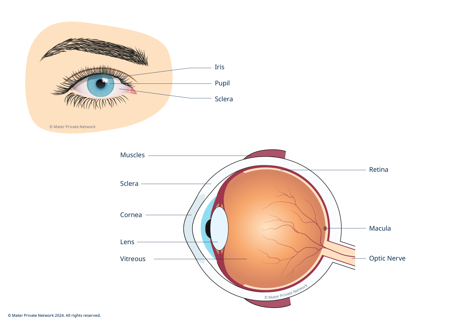
A retinal vein occlusion occurs when small veins in the retina become blocked. The blockage can damage the blood vessels of the retina, the layer of tissue at the back of the inner eye that converts light images to nerve signals and sends them to the brain.
There are two types of retinal vein occlusion:
- Branch retinal vein occlusion (BRVO) affecting a branch of the central retinal vein
- Central retinal vein occlusion (CRVO) affecting the central retinal vein
There are a number of causes of retinal vein occlusion and these can include:
- High blood pressure
- Diabetes
- Blood disorders
- Glaucoma
- Trauma
- Detection

An optician or GP can detect the signs of retinal vein occlusion (RVO). If a RVO is suspected you will be referred to a retinal consultant specialist who has a number of tests or examinations that can be used for diagnosis:
- Visual acuity: measures how well you see at various distances
- Slit lamp examination: examines your eye under high magnification
- Tonometry: measures the pressure in the eye
- Eye examination: examines the retina in detail, using eye drops to dilate the pupil
- Amsler grid: using a lined grid to check for distorted or missing lines
- Colour fundus photography: photography of the fundus (inner lining of the eye), which produces a detailed photograph of the retina and the blood vessels
- Fluorescein angiogram: photography of the blood vessels in the retina using a special dye
- Optical coherence tomography: 3D scans of the layers of the macula part of the retina
There are treatment options to manage RVO and the resulting complications
- Macular oedema - fluid swelling in the macula
- Nacular ischaemia - lack of blood supply
- Neovascularisation - abnormal growth of new blood vessels
Injections
Injections are used to treat swelling of the macula due blood or fluid. The goal of treatment is to prevent further loss of vision. Although some patients have regained vision, the medication may not restore vision that has already been lost and may not ultimately prevent further loss of vision caused by the disease.
Steroid injections
Ozurdex is a biodegradable implant containing the corticosteroid dexamethasone. Corticosteroids, such as dexamethasone, block chemical pathways that lead to inflammation, leakage from the retinal blood vessels and oedema (swelling) of the retina. Two to three injections a year may be required.
Laser therapy
In branch retinal vein occlusion laser photocoagulation is a proven therapy for new blood vessels in vein occlusions. Laser treatment can cause stabilisation or, at times, regression of the new blood vessels.
This treatment, while important in helping to prevent further visual loss, is not usually associated with improvement in vision.
Depending on the treatment you receive, your consultant will advise you on any follow-up care required or ant post-treatment instructions.
The retinal department at Mater Private's eye centre works as a multidisciplinary team delivering treatments and surgery to 5,000 patients annually with routine and complex eye conditions. You are in good hands.


